
Estrogen's Role in the Follicular Phase Explained
Nourished · August 11, 2026
Estrogen’s Role in the Follicular Phase Explained
Estradiol, the most biologically active form of estrogen, drives two processes that make ovulation possible: it thickens the uterine lining and orchestrates a hormonal feedback sequence that ends in the LH surge. According to the Cleveland Clinic, this dual role makes estrogen the central hormone of the follicular phase, not merely a supporting player.
Key entities involved:
- Estradiol (E2): the primary estrogen produced by granulosa cells in growing follicles
- FSH and LH: pituitary gonadotropins that regulate follicle recruitment and trigger ovulation
- Inhibin B: a granulosa-cell peptide that fine-tunes FSH suppression
- NCBI Bookshelf and Cleveland Clinic: primary clinical references for cycle physiology
The follicular phase spans roughly days 1–14 of a typical 28-day cycle, though its length is the most variable segment of any cycle. What follows covers the estradiol timeline from early to late follicular, the cellular machinery behind estrogen production, endometrial changes, neuroendocrine feedback, lab testing, symptoms, fertility implications, and nutrition strategies.
Key Takeaways
Estrogen’s role in the follicular phase is both structural and regulatory: it builds the uterine lining while coordinating the hormonal cascade that makes ovulation possible.
| Point | Details |
|---|---|
| Estradiol drives two core processes | It thickens the endometrium and orchestrates the LH surge that triggers ovulation. |
| E2 rises across three phases | Early follicular: 30–100 pg/mL; mid-follicular: 30–100 pg/mL; late follicular peak: 100–400 pg/mL. |
| Two-cell model produces estrogen | LH stimulates theca androgens; FSH drives granulosa aromatase to convert them to estradiol. |
| Day-3 FSH + E2 + AMH is the standard panel | Pair all three on days 2–4 and add ultrasound when results are borderline or discordant. |
| Nourished supports follicular-phase nutrition | Cycle-aware meal plans address the specific iron, zinc, protein, and fat needs of the follicular phase. |
Table of Contents
- How does estrogen change across the follicular phase?
- How do theca and granulosa cells produce estrogen?
- What does rising estrogen do to the uterus?
- How does estrogen shift pituitary feedback to trigger the LH surge?
- What lab tests measure follicular-phase estrogen?
- What physical and mood changes come with rising estrogen?
- How does follicular-phase estrogen affect fertility timing?
- What causes low or high follicular-phase estrogen?
- What nutrition and lifestyle choices support healthy follicular-phase estrogen?
- A clinician’s perspective on follicular-phase estrogen in practice
- Personalized nutrition built around your follicular phase
- Sources
How does estrogen change across the follicular phase?
The follicular phase divides cleanly into three hormonal windows, each with a distinct estradiol pattern.
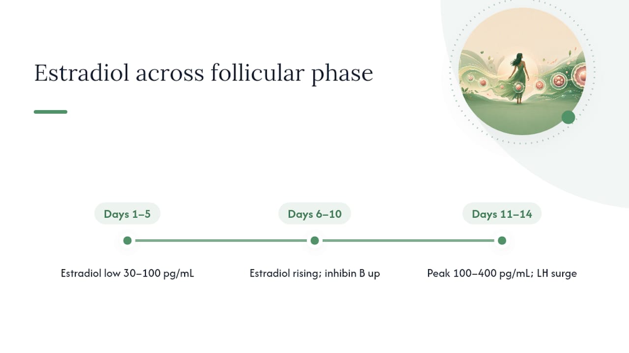
Early follicular (days 1–5): Estradiol sits at its lowest point, typically in the range of 30–100 pg/mL. FSH rises because the corpus luteum from the previous cycle has broken down, and falling progesterone and estrogen release the pituitary from negative feedback. That FSH rise recruits a cohort of antral follicles.
Mid-follicular (days 6–10): As granulosa cells in the recruited follicles proliferate and ramp up aromatase activity, estradiol climbs steadily. One follicle gains a growth and receptor advantage over the others. Estradiol and inhibin B begin suppressing FSH, which starves subordinate follicles of the signal they need to survive. According to StatPearls, this FSH suppression by rising estradiol and inhibin B is the mechanism that times the LH surge and narrows the field to a single dominant follicle.
Late follicular/preovulatory (days 11–14): Estradiol reaches its cycle peak, typically 100–400 pg/mL, and sustains that level for roughly 36–48 hours. That sustained elevation flips pituitary feedback from negative to positive, producing the LH surge and triggering ovulation. The Cleveland Clinic notes that the follicular phase is the most variable segment of the cycle, commonly ranging from 14–21 days, which matters enormously for anyone using cycle-day counting alone to time fertility decisions.
Ranges from Cleveland Clinic estrogen reference data; values vary by lab and assay.
Pro Tip: If your cycle is consistently shorter or longer than 28 days, shift these day-number windows proportionally. A 35-day cycle likely has a longer follicular phase, not a longer luteal phase — the luteal phase stays relatively fixed at around 14 days.
How do theca and granulosa cells produce estrogen?
Estrogen production in the follicle is a two-cell operation. Neither theca cells nor granulosa cells can make estradiol alone. According to Biology LibreTexts, LH stimulates theca cells to synthesize androgens (primarily androstenedione and testosterone), which then diffuse across the basement membrane into granulosa cells. FSH simultaneously stimulates granulosa cells to express aromatase, the enzyme that converts those androgens into estradiol.
The two-cell, two-gonadotropin model means that a signaling failure at either step — abnormal LH pulses, FSH receptor dysfunction, or impaired aromatase activity — can produce low estradiol even when the ovaries appear structurally normal on ultrasound. This is why a blood test showing low E2 is not automatically a diagnosis of ovarian failure; it can reflect a signaling problem upstream.
The sequence from follicle recruitment to dominance follows a clear progression:
- Recruitment: Rising FSH at the start of the cycle activates a cohort of antral follicles that have matured enough to express FSH receptors.
- Growth: FSH drives granulosa cell proliferation and increases aromatase expression, so estradiol output rises in proportion to follicle size.
- Estradiol production: Theca cells supply androgens via LH stimulation; granulosa aromatase converts them to E2, which enters circulation.
- Receptor changes: The dominant follicle acquires LH receptors on its granulosa cells earlier than subordinate follicles, giving it a survival advantage as FSH falls.
- Dominance: The leading follicle sustains estradiol production despite declining FSH; subordinate follicles, unable to maintain growth without adequate FSH, undergo atresia.
- Atresia of subordinates: Apoptosis clears the field, leaving one follicle to complete maturation and ovulate.
Understanding this cascade also explains why disorders like hormonal changes at menarche can disrupt early follicular development — the same FSH/LH signaling axis that governs adult cycles is still being calibrated in adolescence.
What does rising estrogen do to the uterus?
The endometrium during the follicular phase is called the proliferative phase endometrium, and the name is literal. Estradiol drives mitosis in both the epithelial cells lining the uterine cavity and the stromal cells beneath them. Simultaneously, it stimulates angiogenesis through vascular endothelial growth factor (VEGF), building a denser capillary network that will eventually support an implanting embryo. According to NCBI Bookshelf’s reproductive physiology overview, these uterine changes are synchronized with ovarian follicular development through circulating estradiol.
The uterine changes track the ovarian timeline closely:
- Early proliferative (days 1–5): The endometrium is thin, typically 2–4 mm. Glands are narrow and straight. Estradiol is low, and mitotic activity is minimal.
- Mid-proliferative (days 6–10): Glands lengthen and begin to coil. Stromal cells proliferate. The endometrium thickens to roughly 6–8 mm on ultrasound.
- Late proliferative (days 11–14): Glandular coiling increases further. The endometrium reaches 8–12 mm, with a characteristic trilaminar appearance on ultrasound that clinicians use as a marker of adequate estrogen exposure.
Why does this matter beyond fertility? Because the secretory transformation that follows ovulation, driven by progesterone from the corpus luteum, can only occur on an endometrium that estrogen has already prepared. Inadequate estrogen exposure during the follicular phase means progesterone has less to work with, which can impair implantation even when ovulation occurs normally.
How does estrogen shift pituitary feedback to trigger the LH surge?
For most of the follicular phase, estradiol suppresses FSH. This is negative feedback: rising E2 signals the hypothalamus and pituitary to reduce GnRH pulse amplitude and FSH secretion. Inhibin B, secreted by granulosa cells, adds a parallel suppressive signal specifically targeting FSH. The result is a narrowing of follicular competition that allows one dominant follicle to emerge.
The switch to positive feedback is one of the more counterintuitive events in reproductive endocrinology. When estradiol reaches a sustained high level for approximately 36–48 hours, the pituitary’s response flips: instead of suppressing LH, high estradiol now amplifies it. The hypothalamus increases GnRH pulse frequency, and the pituitary becomes hypersensitive to each pulse, releasing a large bolus of LH.
The downstream events after the LH surge unfold rapidly:
- Resumption of meiosis: The oocyte, arrested in prophase I since fetal development, resumes meiotic division in response to LH.
- Matrix metalloproteinase activation: LH triggers proteolytic enzymes that begin digesting the follicle wall.
- Follicle rupture: Prostaglandins and smooth muscle contractions in the follicle wall complete the rupture, releasing the oocyte and its surrounding cumulus cells approximately 36 hours after the LH surge peak.
According to NCBI Bookshelf, the transition from negative to positive feedback is concentration- and duration-dependent, which is why a brief estradiol spike does not trigger an LH surge but a sustained elevation does.
What lab tests measure follicular-phase estrogen?
Hormone testing during the follicular phase gives clinicians a snapshot of ovarian function, but the timing and combination of tests matter as much as the numbers themselves.
Day-3 testing is the standard entry point for ovarian reserve evaluation. On cycle day 3 (or days 2–4), FSH and estradiol are drawn together. A high FSH (generally above 10 IU/L, though lab cutoffs vary) suggests diminished ovarian reserve. Estradiol on day 3 is typically below 80 pg/mL; an elevated day-3 E2 can suppress FSH artificially and make FSH look falsely normal, which is why both values must be interpreted together. StatPearls confirms that day-3 FSH and E2 remain standard tools for assessing ovarian reserve and cycle health.
| Test | Timing | Typical reference range | Clinical interpretation |
|---|---|---|---|
| FSH (day 3) | Days 2–4 | 3–10 IU/L | Elevated FSH suggests reduced reserve |
| E2 (day 3) | Days 2–4 | Below 80 pg/mL | Elevated E2 may suppress FSH; interpret together |
| E2 (mid-follicular) | Days 6–10 | 30–100 pg/mL | Confirms follicular development is progressing |
| E2 (preovulatory peak) | Days 11–14 | 100–400 pg/mL | Sustained peak triggers LH surge |
| AMH | Any cycle day | Lab-specific | Reflects antral follicle count; not cycle-day dependent |
Reference ranges from Medscape and Cleveland Clinic; values vary by assay and laboratory.
AMH (anti-Müllerian hormone) can be drawn on any cycle day, making it a convenient complement to day-3 testing. It reflects the size of the antral follicle pool and does not fluctuate with the acute hormonal changes of the cycle. Inhibin B, while less commonly ordered in routine practice, peaks in the early-to-mid follicular phase and provides additional information about granulosa cell function.
One consistent caveat across all estradiol measurements: assay platforms differ, and a value from one lab is not directly comparable to the same number from another. Medscape’s estradiol reference guide emphasizes interpreting E2 with assay-specific reference ranges rather than universal cutoffs.
Pro Tip: When ordering a fertility hormone panel, pair FSH + E2 + AMH on the same day-3 draw. A single test in isolation rarely tells the full story. If results are borderline, a transvaginal ultrasound antral follicle count adds structural context that blood tests alone cannot provide.
What physical and mood changes come with rising estrogen?
The follicular phase is often described as the “high-energy” phase of the cycle, and the physiology backs that up. As estradiol rises, it interacts with receptors throughout the brain and body, producing changes that many people notice without connecting them to their cycle.
Common signs of rising estrogen during the follicular phase include:
- Improved mood and mental clarity: Estradiol modulates serotonin and dopamine activity, which tends to lift mood and sharpen focus in the mid-follicular phase.
- Increased energy and motivation: Many people report feeling more physically capable and socially engaged as E2 climbs.
- Clearer skin: Estradiol supports collagen synthesis and sebum regulation, often producing a visible improvement in skin texture and tone.
- Changes in cervical mucus: As estradiol rises, cervical mucus becomes more abundant, clear, and stretchy (the “egg white” consistency), a reliable fertility sign.
- Heightened libido: Estrogen’s effect on vaginal lubrication and central arousal pathways often increases sexual interest in the late follicular phase.
The Cleveland Clinic notes that rising estrogen is commonly linked to these physical and mental shifts, and many people use them for wellness or activity planning. Individual variability is real, though. Someone with a short follicular phase may experience these changes compressed into a few days; someone approaching perimenopause may notice them less distinctly as estradiol peaks become lower and less sustained.
Pro Tip: Track cervical mucus alongside a simple daily energy and mood rating (1–5 scale) for two or three cycles. The pattern that emerges is often more informative than a single hormone test, and it gives you a baseline to compare against if symptoms shift.
How does follicular-phase estrogen affect fertility timing?
The fertile window is narrower than most people expect: roughly five days before ovulation and the day of ovulation itself. Estradiol’s rise is the engine behind that window, because it drives both the endometrial preparation and the LH surge that releases the egg.
For natural conception, the practical sequence looks like this:
- Track the estradiol-driven mucus change as the first sign that the late follicular phase is approaching.
- Use an LH urine test to detect the surge, which typically peaks 24–36 hours before ovulation.
- Time intercourse on the day of the LH surge and the day before, when sperm survival and egg viability overlap most reliably.
- Confirm ovulation with a temperature shift (basal body temperature rises 0.2–0.5°C after ovulation) or a post-ovulatory progesterone test.
In assisted reproduction, clinicians use serial serum E2 measurements alongside transvaginal ultrasound to monitor follicular development during IUI or IVF stimulation cycles. A rising E2 paired with a follicle growing roughly 1–2 mm per day toward a target diameter of 18–20 mm signals healthy development. StatPearls and clinical practice guidelines recommend pairing hormone measures with ultrasound because single-point blood values are context-dependent.
Home LH kits are appropriate for most people tracking natural cycles. Serum LH and E2 monitoring becomes necessary when cycles are irregular, when LH kit results are confusing (some people with PCOS have persistently elevated LH), or during medicated cycles where the timing of trigger injections depends on precise E2 and follicle-size thresholds.
Pro Tip: If your E2 is rising but your dominant follicle is smaller than expected for that estradiol level, or if the LH surge is delayed beyond day 16–17, mention both findings to your clinician together. Discordant results are more informative than either value alone and often point to specific management adjustments.
What causes low or high follicular-phase estrogen?
Abnormal estradiol during the follicular phase is rarely a standalone finding. It usually reflects something happening upstream in the hypothalamic-pituitary-ovarian axis or within the follicle itself.
Low follicular estradiol is more common and has several distinct causes:
- Diminished ovarian reserve: Fewer antral follicles means less granulosa cell mass and lower estradiol output, often accompanied by elevated FSH.
- Hypogonadotropic hypogonadism: Low FSH and LH (from hypothalamic suppression due to extreme exercise, low body weight, or chronic stress) means follicles are not adequately stimulated.
- Recent hormonal suppression: Stopping hormonal contraception can temporarily blunt the pituitary-ovarian axis; cycles may take several months to normalize.
- Signaling pathway dysfunction: As Biology LibreTexts explains, aromatase deficiency or FSH/LH receptor mutations can produce low E2 despite normal-appearing ovaries.
- Lab or timing error: A day-3 draw taken on the wrong cycle day, or an assay calibration issue, can produce a spuriously low result worth repeating.
High follicular estradiol is less common in natural cycles but worth recognizing:
- Exogenous estrogen: Hormone therapy or certain supplements can elevate E2 and suppress FSH, mimicking the mid-follicular state.
- PCOS patterns: Polycystic ovary syndrome often disrupts the normal follicular-phase trajectory. The LH/FSH ratio is frequently elevated, follicles recruit but fail to select a dominant one, and estradiol may plateau rather than peak cleanly. Anovulation is the result.
- Estrogen-secreting tumors: Rare, but granulosa cell tumors of the ovary can produce markedly elevated E2 independent of the normal cycle.
Red flags that warrant prompt evaluation: E2 above 80 pg/mL on day 3 (especially with a normal or low FSH), no LH surge detected after day 18–20, or estradiol that fails to rise at all across a full follicular phase. Any of these patterns, combined with absent or irregular periods, should prompt a conversation with a reproductive endocrinologist or gynecologist.
What nutrition and lifestyle choices support healthy follicular-phase estrogen?
Estradiol production requires adequate substrate. The follicle’s theca and granulosa cells need cholesterol (the precursor to all steroid hormones), specific micronutrients, and a hormonal environment that is not chronically suppressed by energy deficit or excess cortisol.
A few evidence-linked levers worth knowing:
- Avoid severe calorie restriction. Hypothalamic suppression from chronic energy deficit is one of the most common causes of blunted follicular-phase estradiol in otherwise healthy people. The hypothalamus reads low energy availability as a signal to downregulate reproductive function.
- Prioritize dietary fat. Steroidogenesis depends on cholesterol. Diets very low in fat, particularly those cutting saturated and monounsaturated fats, can reduce substrate availability for estrogen synthesis.
- Support iron and zinc intake. Iron supports red blood cell production lost during menstruation; zinc is a cofactor in multiple enzymatic steps of steroidogenesis. Both are commonly low in people with heavy periods or restrictive diets.
- Match exercise intensity to phase. High-intensity training is generally well-tolerated in the follicular phase when estradiol is rising, but chronic overtraining without adequate recovery suppresses GnRH pulsatility. The follicular phase is a good window for progressive loading; the key is recovery nutrition, particularly protein intake, which supports both muscle repair and hormone synthesis.
- Manage sleep and cortisol. Cortisol competes with progesterone for the same receptor and can blunt the hypothalamic GnRH signal. Consistent sleep (7–9 hours) and stress management are not optional extras for cycle health.
When symptoms or test results suggest a hormonal pattern worth addressing, a registered dietitian with reproductive health experience can translate these principles into a practical eating plan. Nourished’s cycle-aware nutrition platform builds meal plans around phase-specific needs, including the higher energy demands and micronutrient priorities of the follicular phase.
Pro Tip: For two cycles, log food intake, sleep hours, and exercise intensity alongside your cycle day. Patterns often emerge quickly: a cluster of short follicular phases may correlate with low-calorie weeks, or delayed ovulation with high-stress periods. That data is far more useful to a clinician than a single hormone panel.
A clinician’s perspective on follicular-phase estrogen in practice
The physiology covered in this article is not just academic. Clinicians use it daily to make concrete decisions, and understanding it changes how you engage with your own care.
When a reproductive endocrinologist orders a day-3 FSH and E2, they are not just checking boxes. They are asking: is the pituitary working appropriately hard to recruit follicles, and is the ovary responding with estradiol? A high FSH with a low E2 tells a different story than a high FSH with a high E2, even though both involve an elevated FSH. The E2 value contextualizes the FSH, and AMH contextualizes both. Ordering all three together is standard practice for a reason.
Nutritionists working with cycle-aware frameworks use the follicular phase differently than the luteal phase. The rising estradiol of the follicular phase supports higher energy availability, better insulin sensitivity, and faster recovery from exercise. That makes it the right window for higher-intensity training, progressive dietary changes, and introducing new foods or eating patterns. Trying to make the same changes in the late luteal phase, when progesterone dominates and appetite and fatigue shift, tends to produce worse adherence.
One practical recommendation worth taking to your next appointment: if you have had a hormone panel done but only received a single number without context (cycle day, assay platform, paired tests), ask for the full picture. A day-3 E2 of 60 pg/mL is reassuring. The same number drawn on day 8 of a 30-day cycle is a different conversation entirely.
Personalized nutrition built around your follicular phase
The physiology of the follicular phase is precise. Your nutrition plan should be too.
Nourished builds personalized, cycle-aware meal plans that adapt to where you are in your cycle, including the specific energy, protein, and micronutrient priorities of the follicular phase. Rather than a generic eating template, you get a plan that shifts with your hormones.
- Phase-specific meals: Follicular-phase plans emphasize iron, zinc, and adequate dietary fat to support estrogen synthesis and recovery from menstruation.
- Lab and symptom tracking compatibility: Log your E2, FSH, or AMH results alongside food and energy data to see how nutrition patterns correlate with your cycle.
- Evidence-backed resources: Nourished’s research references underpin every recommendation, so you know the guidance connects to real reproductive physiology.
Ready to eat in sync with your cycle? Start your personalized plan at Nourished.
Sources
- Follicular Phase of Menstrual Cycle — Cleveland Clinic
- The Normal Menstrual Cycle and the Control of Ovulation — NCBI Bookshelf
- 25.4 Female Reproductive Physiology — Biology LibreTexts
- Estradiol: Reference Range, Interpretation, Collection and … — Medscape
- The Normal Menstrual Cycle and the Control of Ovulation — Endotext (PDF)
This article provides general health information and is not a substitute for professional medical advice. Consult a qualified clinician or reproductive endocrinologist for evaluation and treatment of hormonal concerns.